Virtual cellular pathology laboratory suite
Immersive laboratory experience with realistic workflows, from specimen processing to advanced molecular techniques. Develop the practical skills and clinical confidence interview panels probe at every band.
Virtual Cellular Pathology Laboratory Suite is available to premium subscribers and trial users.
Cellular pathology result interpretation module
AI-assisted histopathology report interpretation following UK best practice: RCPath Datasets, NICE Guidelines, UICC TNM 9 staging and IBMS competency frameworks.
Cellular Pathology Result Interpretation Module is available to premium subscribers and trial users.
Cellular pathology LIMS system
Advanced laboratory information management for cellular pathology with cross-department references, specimen tracking and validation tools.
Cellular Pathology LIMS System is available to premium subscribers and trial users.
Histological staining methods explorer
Explore different staining techniques, their applications and interpretation guidelines for enhanced diagnostic accuracy.
Staining Methods Explorer
Explore histological staining techniques and their applications in diagnostic pathology
Staining Techniques
Select a staining method to view details
Understanding staining methods is critical for accurate histopathological diagnosis
Tissue pattern recognition
Master the identification of key histological patterns essential for accurate diagnostics and patient care.
Tissue Pattern Recognition
Master key histological patterns for accurate diagnosis and interpretation
Histological Patterns
Granulomatous Inflammation
Characterised by aggregates of epithelioid histiocytes, often with multinucleated giant cells and surrounding lymphocytes.
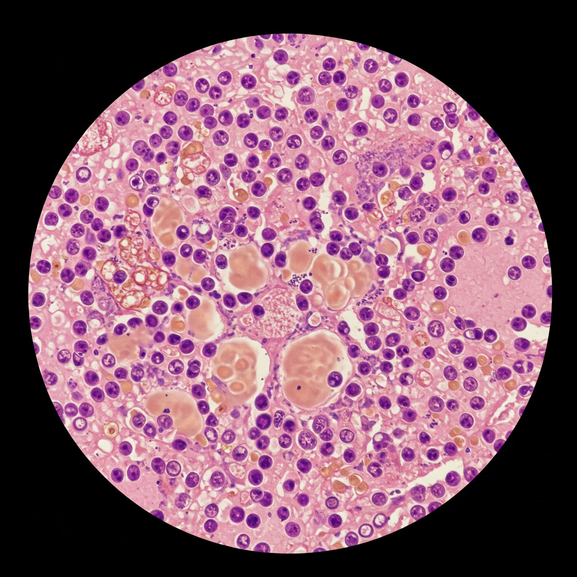
Granulomatous Inflammation
Characterised by aggregates of epithelioid histiocytes, often with multinucleated giant cells and surrounding lymphocytes.
Key Characteristics
- Epithelioid histiocytes with abundant pale cytoplasm
- Multinucleated giant cells (Langhans or foreign body type)
- Lymphocytic rim surrounding granulomas
- Variable necrosis (caseating vs. non-caseating)
Pattern recognition is a fundamental skill for all histopathologists and biomedical scientists
Cellular pathology data visualisation
Interactive charts and graphs showcasing key cellular pathology metrics, staining techniques and their clinical significance.
Histological Staining Techniques
Comparative usage and diagnostic value of different staining methods in clinical histopathology
Abbreviations:
- H&E: Haematoxylin and Eosin
- IHC: Immunohistochemistry
- ISH: In Situ Hybridization
- FISH: Fluorescence In Situ Hybridization
Cellular pathology quick guide
Essential knowledge areas for cellular pathology, from foundational concepts to specialised techniques.
Tissue Processing
Fixation methods, embedding techniques, sectioning, and staining procedures for optimal tissue preservation and visualisation.
Staining Techniques
Special stains, H&E, immunohistochemistry methods, and their applications in different diagnostic contexts.
Microscopy
Light microscopy, electron microscopy, fluorescence techniques, and digital imaging systems used in modern histology.
Molecular Techniques
In-situ hybridization, PCR applications, next-generation sequencing, and their integration with traditional histology.
Digital Pathology
Whole slide imaging, image analysis algorithms, AI applications, and implementation of digital workflows.
Quality Management
Internal quality control, external quality assessment, accreditation standards, and continuous improvement processes.
Sources and guidance
This hub's content aligns with the UK's professional and regulatory references for the specialty: